Circulatory System and Respiratory System Test Review Answers
Chapter eleven: Introduction to the Torso's Systems
11.3 Circulatory and Respiratory Systems
Learning Objectives
Past the cease of this section, you will be able to:
- Draw the passage of air from the outside environment to the lungs
- Explicate how the lungs are protected from particulate matter
- Describe the part of the circulatory organization
- Depict the cardiac cycle
- Explain how blood flows through the body
Animals are circuitous multicellular organisms that require a machinery for transporting nutrients throughout their bodies and removing wastes. The homo circulatory system has a circuitous network of blood vessels that reach all parts of the torso. This extensive network supplies the cells, tissues, and organs with oxygen and nutrients, and removes carbon dioxide and waste compounds.
The medium for transport of gases and other molecules is the blood, which continually circulates through the organisation. Pressure differences within the system cause the movement of the blood and are created by the pumping of the heart.
Gas exchange between tissues and the blood is an essential function of the circulatory organization. In humans, other mammals, and birds, blood absorbs oxygen and releases carbon dioxide in the lungs. Thus the circulatory and respiratory system, whose part is to obtain oxygen and discharge carbon dioxide, piece of work in tandem.
The Respiratory System (Basic level)
Take a breath in and hold it. Look several seconds and and then permit it out. Humans, when they are not exerting themselves, breathe approximately 15 times per minute on average. This equates to near 900 breaths an 60 minutes or 21,600 breaths per twenty-four hour period. With every inhalation, air fills the lungs, and with every exhalation, information technology rushes back out. That air is doing more than than just inflating and deflating the lungs in the chest crenel. The air contains oxygen that crosses the lung tissue, enters the bloodstream, and travels to organs and tissues. There, oxygen is exchanged for carbon dioxide, which is a cellular waste material. Carbon dioxide exits the cells, enters the bloodstream, travels dorsum to the lungs, and is expired out of the body during exhalation.
Animate is both a voluntary and an involuntary event. How often a jiff is taken and how much air is inhaled or exhaled is regulated by the respiratory center in the brain in response to signals it receives nearly the carbon dioxide content of the blood. Notwithstanding, information technology is possible to override this automatic regulation for activities such as speaking, singing and pond under water.
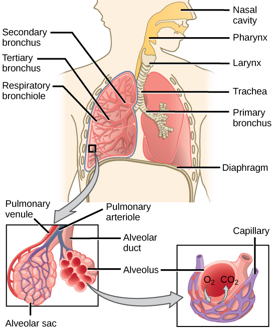
During inhalation the diaphragm descends creating a negative pressure around the lungs and they begin to inflate, drawing in air from exterior the trunk. The air enters the trunk through the nasal cavity located just inside the olfactory organ (Figure 11.9). As the air passes through the nasal crenel, the air is warmed to torso temperature and humidified by wet from mucous membranes. These processes help equilibrate the air to the body conditions, reducing any impairment that cold, dry air can cause. Particulate matter that is floating in the air is removed in the nasal passages by hairs, mucus, and cilia. Air is also chemically sampled by the sense of smell.
From the nasal cavity, air passes through the pharynx (pharynx) and the larynx (vox box) equally it makes its way to the trachea (Effigy 11.nine). The chief part of the trachea is to funnel the inhaled air to the lungs and the exhaled air dorsum out of the body. The human trachea is a cylinder, well-nigh 25 to 30 cm (ix.viii–xi.8 in) long, which sits in front of the esophagus and extends from the pharynx into the chest cavity to the lungs. Information technology is made of incomplete rings of cartilage and smoothen muscle. The cartilage provides strength and back up to the trachea to keep the passage open. The trachea is lined with cells that have cilia and secrete mucus. The mucus catches particles that have been inhaled, and the cilia move the particles toward the pharynx.
The end of the trachea divides into two bronchi that enter the right and left lung. Air enters the lungs through the primary bronchi. The primary bronchus divides, creating smaller and smaller diameter bronchi until the passages are under 1 mm (.03 in) in diameter when they are called bronchioles equally they carve up and spread through the lung. Like the trachea, the bronchus and bronchioles are made of cartilage and smooth muscle. Bronchi are innervated by nerves of both the parasympathetic and sympathetic nervous systems that command muscle contraction (parasympathetic) or relaxation (sympathetic) in the bronchi and bronchioles, depending on the nervous system's cues. The final bronchioles are the respiratory bronchioles. Alveolar ducts are attached to the end of each respiratory bronchiole. At the stop of each duct are alveolar sacs, each containing 20 to thirty alveoli. Gas exchange occurs only in the alveoli. The alveoli are thin-walled and look like tiny bubbles within the sacs. The alveoli are in straight contact with capillaries of the circulatory system. Such intimate contact ensures that oxygen will diffuse from the alveoli into the claret. In addition, carbon dioxide will lengthened from the blood into the alveoli to be exhaled. The anatomical arrangement of capillaries and alveoli emphasizes the structural and functional human relationship of the respiratory and circulatory systems. Estimates for the expanse of alveoli in the lungs vary around 100 thoutwo. This large area is nigh the expanse of half a lawn tennis court. This big surface expanse, combined with the thin-walled nature of the alveolar cells, allows gases to hands diffuse beyond the cells.
Systems of Gas Exchange
The main role of the respiratory system is to deliver oxygen to the cells of the torso's tissues and remove carbon dioxide, a prison cell waste production. The chief structures of the homo respiratory system are the nasal cavity, the trachea, and lungs.
All aerobic organisms require oxygen to comport out their metabolic functions. Along the evolutionary tree, different organisms have devised different ways of obtaining oxygen from the surrounding atmosphere. The environment in which the animal lives profoundly determines how an fauna respires. The complication of the respiratory system is correlated with the size of the organism. Equally animal size increases, improvidence distances increment and the ratio of surface surface area to volume drops. In unicellular organisms, improvidence across the prison cell membrane is sufficient for supplying oxygen to the prison cell (Figure 11.ten). Diffusion is a slow, passive transport procedure. In order for diffusion to be a feasible means of providing oxygen to the cell, the rate of oxygen uptake must match the charge per unit of diffusion across the membrane. In other words, if the cell were very large or thick, diffusion would not exist able to provide oxygen apace enough to the inside of the cell. Therefore, dependence on improvidence every bit a means of obtaining oxygen and removing carbon dioxide remains feasible but for small organisms or those with highly-flattened bodies, such as many flatworms (Platyhelminthes). Larger organisms had to evolve specialized respiratory tissues, such as gills, lungs, and respiratory passages accompanied past a circuitous circulatory systems, to ship oxygen throughout their entire body.
Straight Improvidence
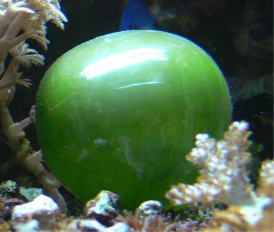
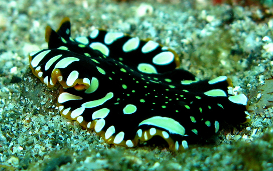
For small-scale multicellular organisms, diffusion beyond the outer membrane is sufficient to meet their oxygen needs. Gas commutation by direct diffusion across surface membranes is efficient for organisms less than i mm in diameter. In simple organisms, such as cnidarians and flatworms, every cell in the trunk is close to the external environment. Their cells are kept moist and gases diffuse quickly via directly diffusion. Flatworms are pocket-sized, literally apartment worms, which 'breathe' through diffusion beyond the outer membrane (Figure eleven.xi). The flat shape of these organisms increases the surface area for diffusion, ensuring that each cell inside the trunk is close to the outer membrane surface and has access to oxygen. If the flatworm had a cylindrical trunk, and then the cells in the center would not be able to go oxygen.
Pare and Gills
Earthworms and amphibians utilise their skin (integument) equally a respiratory organ. A dense network of capillaries lies just beneath the skin and facilitates gas exchange between the external environment and the circulatory system. The respiratory surface must be kept moist in order for the gases to dissolve and diffuse beyond cell membranes.
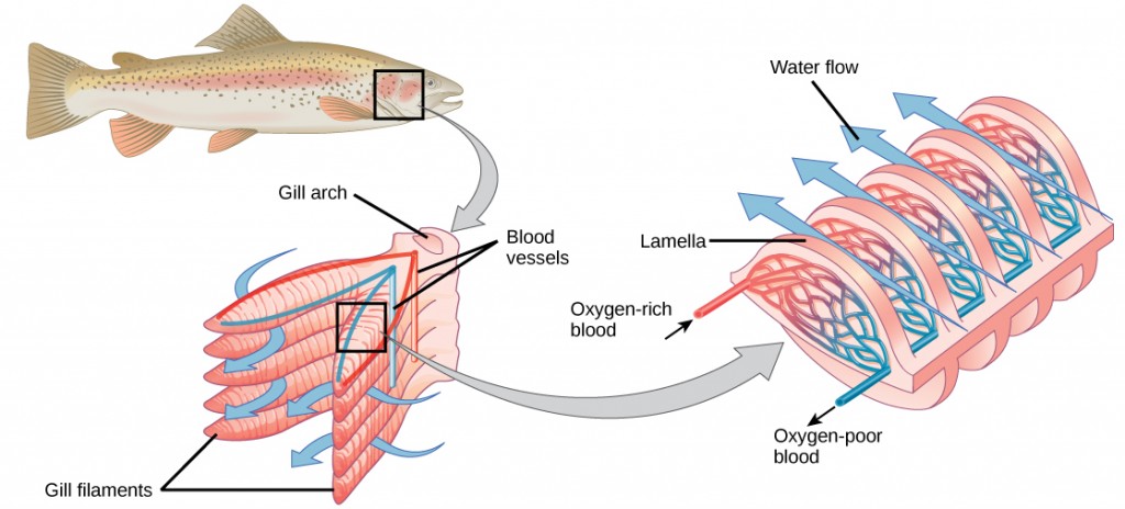
Organisms that live in h2o need to obtain oxygen from the h2o. Oxygen dissolves in water but at a lower concentration than in the atmosphere. The temper has roughly 21 percent oxygen. In water, the oxygen concentration is much smaller than that. Fish and many other aquatic organisms have evolved gills to take up the dissolved oxygen from water (Effigy 11.12). Gills are thin tissue filaments that are highly branched and folded. When water passes over the gills, the dissolved oxygen in water rapidly diffuses beyond the gills into the bloodstream. The circulatory system tin can and then behave the oxygenated blood to the other parts of the body. In animals that incorporate coelomic fluid instead of claret, oxygen diffuses beyond the gill surfaces into the coelomic fluid. Gills are institute in mollusks, annelids, and crustaceans.
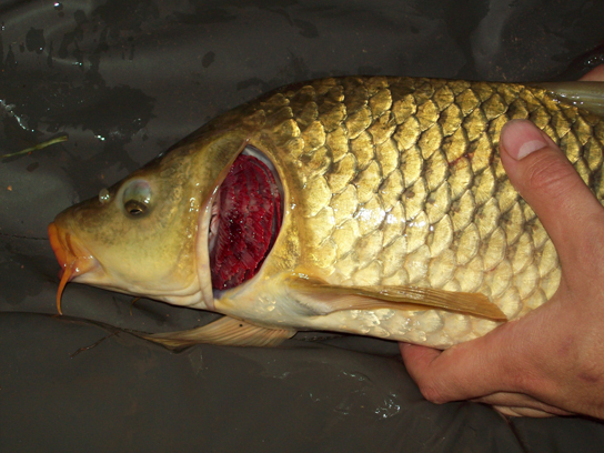
This common carp, like many other aquatic organisms, has gills that let it to obtain oxygen from water. (credit: "Guitardude012″/Wikimedia Commons)
The folded surfaces of the gills provide a large surface expanse to ensure that the fish gets sufficient oxygen. Diffusion is a procedure in which material travels from regions of high concentration to depression concentration until equilibrium is reached. In this case, blood with a low concentration of oxygen molecules circulates through the gills. The concentration of oxygen molecules in h2o is higher than the concentration of oxygen molecules in gills. Every bit a result, oxygen molecules diffuse from water (high concentration) to blood (depression concentration), as shown in Figure 11.13. Similarly, carbon dioxide molecules in the claret diffuse from the blood (high concentration) to water (depression concentration).
Tracheal Systems
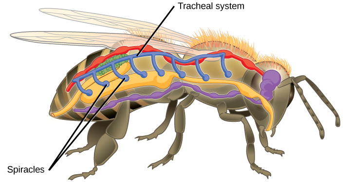
Insect respiration is independent of its circulatory arrangement; therefore, the blood does not play a directly role in oxygen ship. Insects have a highly specialized type of respiratory arrangement called the tracheal system, which consists of a network of small tubes that carries oxygen to the entire trunk. The tracheal system is the most directly and efficient respiratory system in active animals. The tubes in the tracheal organisation are made of a polymeric material chosen chitin.
Insect bodies have openings, called spiracles, forth the thorax and belly. These openings connect to the tubular network, assuasive oxygen to laissez passer into the body (Figure 11.14) and regulating the diffusion of CO2 and water vapor. Air enters and leaves the tracheal arrangement through the spiracles. Some insects tin can ventilate the tracheal organisation with torso movements.
Mammalian Systems
In mammals, pulmonary ventilation occurs via inhalation (animate). During inhalation, air enters the torso through the nasal cavity located just inside the olfactory organ (Figure 11.fifteen). As air passes through the nasal cavity, the air is warmed to torso temperature and humidified. The respiratory tract is coated with mucus to seal the tissues from directly contact with air. Mucus is high in water. As air crosses these surfaces of the mucous membranes, it picks upward h2o. These processes help equilibrate the air to the body atmospheric condition, reducing any harm that cold, dry out air can crusade. Particulate matter that is floating in the air is removed in the nasal passages via mucus and cilia. The processes of warming, humidifying, and removing particles are of import protective mechanisms that preclude harm to the trachea and lungs. Thus, inhalation serves several purposes in add-on to bringing oxygen into the respiratory system.
Which of the following statements about the mammalian respiratory system is false?
- When we breathe in, air travels from the pharynx to the trachea.
- The bronchioles branch into bronchi.
- Alveolar ducts connect to alveolar sacs.
- Gas exchange between the lung and blood takes place in the alveolus.
From the nasal cavity, air passes through the pharynx (throat) and the larynx (voice box), as it makes its fashion to the trachea (Figure 11.16). The chief function of the trachea is to funnel the inhaled air to the lungs and the exhaled air dorsum out of the body. The human trachea is a cylinder about 10 to 12 cm long and 2 cm in bore that sits in front of the esophagus and extends from the larynx into the chest cavity where it divides into the two principal bronchi at the midthorax. Information technology is made of incomplete rings of hyaline cartilage and smooth muscle (Figure eleven.17). The trachea is lined with mucus-producing goblet cells and ciliated epithelia. The cilia propel foreign particles trapped in the fungus toward the pharynx. The cartilage provides strength and support to the trachea to keep the passage open up. The polish muscle tin can contract, decreasing the trachea's bore, which causes expired air to rush upwards from the lungs at a great force. The forced exhalation helps expel fungus when we cough. Smooth muscle can contract or relax, depending on stimuli from the external surround or the torso's nervous system.
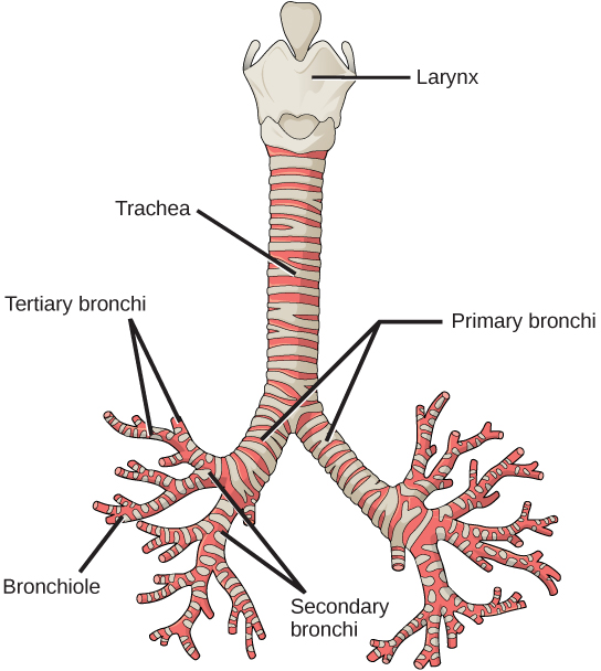
The trachea and bronchi are made of incomplete rings of cartilage. (credit: modification of work by Gray's Anatomy)
Lungs: Bronchi and Alveoli
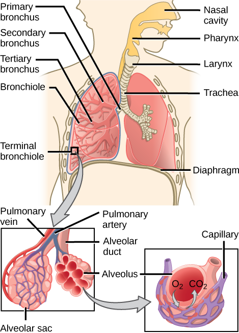
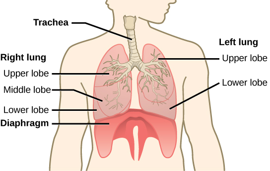
The end of the trachea bifurcates (divides) to the correct and left lungs. The lungs are not identical. The right lung is larger and contains three lobes, whereas the smaller left lung contains two lobes (Effigy eleven.17). The muscular diaphragm, which facilitates animate, is junior (below) to the lungs and marks the terminate of the thoracic cavity.
In the lungs, air is diverted into smaller and smaller passages, or bronchi. Air enters the lungs through the 2 primary (main) bronchi (singular: bronchus). Each bronchus divides into secondary bronchi, then into tertiary bronchi, which in turn split, creating smaller and smaller diameter bronchioles as they split and spread through the lung. Like the trachea, the bronchi are made of cartilage and smooth musculus. At the bronchioles, the cartilage is replaced with elastic fibers. Bronchi are innervated past nerves of both the parasympathetic and sympathetic nervous systems that control musculus contraction (parasympathetic) or relaxation (sympathetic) in the bronchi and bronchioles, depending on the nervous system'south cues. In humans, bronchioles with a diameter smaller than 0.5 mm are the respiratory bronchioles. They lack cartilage and therefore rely on inhaled air to back up their shape. As the passageways decrease in diameter, the relative corporeality of smooth muscle increases.
The terminal bronchioles subdivide into microscopic branches chosen respiratory bronchioles. The respiratory bronchioles subdivide into several alveolar ducts. Numerous alveoli and alveolar sacs surround the alveolar ducts. The alveolar sacs resemble bunches of grapes tethered to the end of the bronchioles (Figure eleven.xviii). In the acinar region, the alveolar ducts are attached to the end of each bronchiole. At the end of each duct are approximately 100 alveolar sacs, each containing 20 to 30 alveoli that are 200 to 300 microns in diameter. Gas substitution occurs only in alveoli. Alveoli are made of sparse-walled parenchymal cells, typically one-cell thick, that await like tiny bubbles within the sacs. Alveoli are in directly contact with capillaries (i-cell thick) of the circulatory organisation. Such intimate contact ensures that oxygen volition diffuse from alveoli into the blood and be distributed to the cells of the body. In addition, the carbon dioxide that was produced by cells as a waste product will lengthened from the blood into alveoli to exist exhaled. The anatomical arrangement of capillaries and alveoli emphasizes the structural and functional relationship of the respiratory and circulatory systems. Because there are so many alveoli (~300 one thousand thousand per lung) within each alveolar sac and so many sacs at the cease of each alveolar duct, the lungs have a sponge-like consistency. This organization produces a very big surface area that is available for gas exchange. The surface area of alveoli in the lungs is approximately 75 m2. This large surface area, combined with the thin-walled nature of the alveolar parenchymal cells, allows gases to easily diffuse beyond the cells.
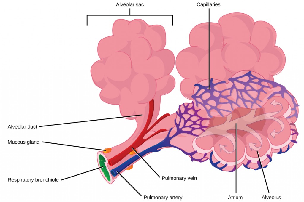
Last bronchioles are continued by respiratory bronchioles to alveolar ducts and alveolar sacs. Each alveolar sac contains 20 to 30 spherical alveoli and has the appearance of a bunch of grapes. Air flows into the atrium of the alveolar sac, and then circulates into alveoli where gas substitution occurs with the capillaries. Mucous glands secrete mucous into the airways, keeping them moist and flexible. (credit: modification of work by Mariana Ruiz Villareal)
Concept in Action

Lookout the following video to review the respiratory organization.
Protective Mechanisms
The air that organisms exhale contains particulate matter such equally dust, dirt, viral particles, and bacteria that tin harm the lungs or trigger allergic immune responses. The respiratory system contains several protective mechanisms to avoid bug or tissue damage. In the nasal crenel, hairs and mucus trap small particles, viruses, bacteria, dust, and clay to foreclose their entry.
If particulates do brand it beyond the olfactory organ, or enter through the oral cavity, the bronchi and bronchioles of the lungs besides contain several protective devices. The lungs produce mucus—a glutinous substance made of mucin, a complex glycoprotein, also as salts and water—that traps particulates. The bronchi and bronchioles contain cilia, pocket-size pilus-similar projections that line the walls of the bronchi and bronchioles (Effigy 11.19). These cilia beat in unison and movement mucus and particles out of the bronchi and bronchioles back up to the throat where it is swallowed and eliminated via the esophagus.
In humans, for example, tar and other substances in cigarette smoke destroy or paralyze the cilia, making the removal of particles more difficult. In addition, smoking causes the lungs to produce more than mucus, which the damaged cilia are non able to motility. This causes a persistent cough, as the lungs try to rid themselves of particulate matter, and makes smokers more susceptible to respiratory ailments.
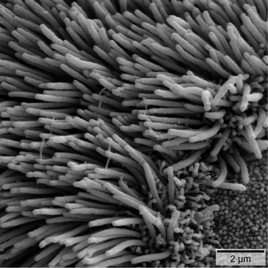
The bronchi and bronchioles contain cilia that help motion mucus and other particles out of the lungs. (credit: Louisa Howard, modification of piece of work by Dartmouth Electron Microscope Facility)
Summary
Animal respiratory systems are designed to facilitate gas exchange. In mammals, air is warmed and humidified in the nasal cavity. Air and then travels down the pharynx, through the trachea, and into the lungs. In the lungs, air passes through the branching bronchi, reaching the respiratory bronchioles, which business firm the first site of gas exchange. The respiratory bronchioles open up into the alveolar ducts, alveolar sacs, and alveoli. Because in that location are and then many alveoli and alveolar sacs in the lung, the expanse for gas exchange is very large. Several protective mechanisms are in place to prevent impairment or infection. These include the hair and fungus in the nasal cavity that trap dust, dirt, and other particulate matter before they tin can enter the system. In the lungs, particles are trapped in a mucus layer and transported via cilia upward to the esophageal opening at the top of the trachea to be swallowed.
The Circulatory System
The circulatory system is a network of vessels—the arteries, veins, and capillaries—and a pump, the heart. In all vertebrate organisms this is a closed-loop system, in which the blood is largely separated from the body's other extracellular fluid compartment, the interstitial fluid, which is the fluid bathing the cells. Blood circulates inside blood vessels and circulates unidirectionally from the heart around one of two circulatory routes, and then returns to the heart again; this is a closed circulatory system. Open circulatory systems are found in invertebrate animals in which the circulatory fluid bathes the internal organs direct even though it may be moved about with a pumping heart.
The Heart
The heart is a circuitous muscle that consists of two pumps: one that pumps blood through pulmonary circulation to the lungs, and the other that pumps blood through systemic circulation to the remainder of the body's tissues (and the eye itself).
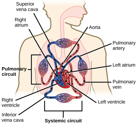
The middle is asymmetrical, with the left side being larger than the correct side, correlating with the different sizes of the pulmonary and systemic circuits (Figure 11.10). In humans, the eye is near the size of a clenched fist; it is divided into four chambers: ii atria and two ventricles. There is one atrium and one ventricle on the right side and ane atrium and one ventricle on the left side. The correct atrium receives deoxygenated claret from the systemic circulation through the major veins: the superior vena cava, which drains blood from the caput and from the veins that come from the artillery, as well as the inferior vena cava, which drains blood from the veins that come from the lower organs and the legs. This deoxygenated blood and so passes to the right ventricle through the tricuspid valve, which prevents the backflow of blood. After information technology is filled, the right ventricle contracts, pumping the blood to the lungs for reoxygenation. The left atrium receives the oxygen-rich blood from the lungs. This claret passes through the bicuspid valve to the left ventricle where the blood is pumped into the aorta. The aorta is the major artery of the torso, taking oxygenated blood to the organs and muscles of the trunk. This pattern of pumping is referred to as double circulation and is establish in all mammals. (Figure 11.twenty).
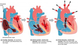
The Cardiac Cycle
The main purpose of the heart is to pump blood through the torso; it does so in a repeating sequence called the cardiac cycle. The cardiac cycle is the flow of blood through the heart coordinated by electrochemical signals that cause the centre muscle to contract and relax. In each cardiac bicycle, a sequence of contractions pushes out the blood, pumping it through the body; this is followed past a relaxation phase, where the heart fills with blood. These two phases are chosen the systole (wrinkle) and diastole (relaxation), respectively (Figure 11.21). The indicate for contraction begins at a location on the outside of the right atrium. The electrochemical bespeak moves from there across the atria causing them to contract. The contraction of the atria forces claret through the valves into the ventricles. Closing of these valves acquired past the contraction of the ventricles produces a "lub" sound. The signal has, by this fourth dimension, passed downwards the walls of the middle, through a point between the right atrium and correct ventricle. The signal then causes the ventricles to contract. The ventricles contract together forcing blood into the aorta and the pulmonary arteries. Closing of the valves to these arteries caused by blood being drawn back toward the middle during ventricular relaxation produces a monosyllabic "dub" sound.
The pumping of the heart is a part of the cardiac muscle cells, or cardiomyocytes, that brand up the middle musculus. Cardiomyocytes are distinctive musculus cells that are striated like skeletal muscle but pump rhythmically and involuntarily like smooth musculus; adjacent cells are connected by intercalated disks plant only in cardiac muscle. These connections allow the electric bespeak to travel directly to neighboring musculus cells.
The electrical impulses in the center produce electrical currents that menstruum through the body and tin can be measured on the skin using electrodes. This data can be observed as an electrocardiogram (ECG) a recording of the electrical impulses of the cardiac musculus.
Concept in Activeness

Visit the post-obit website to see the heart'southward pacemaker, or electrocardiogram system, in action.
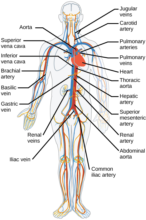
Claret Vessels
The claret from the heart is carried through the body by a circuitous network of claret vessels (Figure 11.22). Arteries have blood away from the heart. The main artery of the systemic circulation is the aorta; it branches into major arteries that accept blood to different limbs and organs. The aorta and arteries near the heart have heavy just rubberband walls that reply to and shine out the pressure differences caused by the chirapsia center. Arteries farther away from the heart have more muscle tissue in their walls that can constrict to touch on flow rates of blood. The major arteries diverge into small-scale arteries, and so smaller vessels called arterioles, to achieve more than deeply into the muscles and organs of the trunk.
Arterioles diverge into capillary beds. Capillary beds contain a large number, x's to 100's of capillaries that branch among the cells of the torso. Capillaries are narrow-diameter tubes that can fit single red blood cells and are the sites for the exchange of nutrients, waste, and oxygen with tissues at the cellular level. Fluid likewise leaks from the blood into the interstitial infinite from the capillaries. The capillaries converge once again into venules that connect to minor veins that finally connect to major veins. Veins are blood vessels that bring blood high in carbon dioxide back to the centre. Veins are not as thick-walled as arteries, since pressure is lower, and they have valves along their length that preclude backflow of claret abroad from the eye. The major veins drain blood from the aforementioned organs and limbs that the major arteries supply.
Section Summary
Animal respiratory systems are designed to facilitate gas substitution. In mammals, air is warmed and humidified in the nasal cavity. Air and so travels down the pharynx and larynx, through the trachea, and into the lungs. In the lungs, air passes through the branching bronchi, reaching the respiratory bronchioles. The respiratory bronchioles open up into the alveolar ducts, alveolar sacs, and alveoli. Because in that location are and so many alveoli and alveolar sacs in the lung, the area for gas exchange is very large.
The mammalian circulatory organisation is a closed arrangement with double circulation passing through the lungs and the torso. It consists of a network of vessels containing blood that circulates because of pressure differences generated past the heart.
The heart contains ii pumps that move blood through the pulmonary and systemic circulations. In that location is one atrium and one ventricle on the right side and ane atrium and one ventricle on the left side. The pumping of the heart is a function of cardiomyocytes, distinctive muscle cells that are striated like skeletal musculus but pump rhythmically and involuntarily like smooth muscle. The signal for wrinkle begins in the wall of the right atrium. The electrochemical signal causes the two atria to contract in unison; then the betoken causes the ventricles to contract. The blood from the heart is carried through the body by a circuitous network of blood vessels; arteries take claret away from the middle, and veins bring blood back to the heart.
Glossary
alveolus: (plural: alveoli) (also, air sacs) the terminal structure of the lung passage where gas commutation occurs
aorta: the major artery that takes blood away from the middle to the systemic circulatory arrangement
artery: a blood vessel that takes claret away from the heart
atrium: (plural: atria) a chamber of the centre that receives blood from the veins
bicuspid valve: a one-mode opening between the atrium and the ventricle in the left side of the eye
bronchi: (singular: bronchus) smaller branches of cartilaginous tissue that stalk off of the trachea; air is funneled through the bronchi to the region where gas exchange occurs in the alveoli
bronchiole: an airway that extends from the main bronchus to the alveolar sac
capillary: the smallest blood vessel that allows the passage of individual claret cells and the site of improvidence of oxygen and nutrient exchange
cardiac cycle: the filling and emptying the centre of blood caused by electrical signals that crusade the heart muscles to contract and relax
closed circulatory arrangement: a system that has the blood separated from the actual interstitial fluid and contained in blood vessels
diaphragm: a skeletal muscle located nether lungs that encloses the lungs in the thorax
diastole: the relaxation phase of the cardiac cycle when the heart is relaxed and the ventricles are filling with blood
electrocardiogram (ECG): a recording of the electric impulses of the cardiac musculus
inferior vena cava: the major vein of the body returning blood from the lower parts of the trunk to the right atrium
larynx: the vocalism box, located within the throat
nasal crenel: an opening of the respiratory system to the exterior environment
open up circulatory system: a circulatory system that has the blood mixed with interstitial fluid in the body crenel and directly bathes the organs
pharynx: the throat
principal bronchus: (also, chief bronchus) a region of the airway within the lung that attaches to the trachea and bifurcates to form the bronchioles
pulmonary circulation: the flow of blood away from the heart through the lungs where oxygenation occurs and and then back to the heart
superior vena cava: the major vein of the body returning claret from the upper part of the body to the right atrium
systemic circulation: the flow of blood away from the eye to the encephalon, liver, kidneys, stomach, and other organs, the limbs, and the muscles of the body, then back to the heart
systole: the contraction stage of cardiac cycle when the ventricles are pumping blood into the arteries
trachea: the cartilaginous tube that transports air from the throat to the lungs
tricuspid valve: a one-fashion opening between the atrium and the ventricle in the right side of the heart
vein: a claret vessel that brings claret back to the heart
ventricle: (of the heart) a large chamber of the heart that pumps claret into arteries
Source: https://opentextbc.ca/biology/chapter/11-3-circulatory-and-respiratory-systems/
Post a Comment for "Circulatory System and Respiratory System Test Review Answers"